
Figure 1. New MENE Mutants Disrupt Internalization from the Cell Surface.
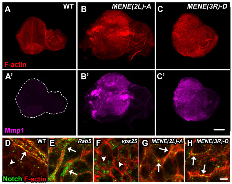
WT (A, A′) or mutant MENE(2L)-A or MENE(3R)-D eye imaginal discs (B, B′ and C, C′) stained for F-actin (red) and Mmp1 (magenta), an indicator of neoplastic transformation. D-H: Eye imaginal discs isolated from wandering L3 larvae stained for Notch (green) and F-actin (red): Notch localizes to the apical plasma membrane and in endocytic vesicles in WT cells (D), in a diffuse subcortical pattern in Rab5 mutants (E), in large internal puncta in Vps25 mutants (F), and predominantly in puncta along the cell cortex in mutants from complementation groups MENE(2L)-A and MENE(3R)-D (G and H). In D-H arrows indicate Notch at the cortex and arrowheads indicate internal Notch. Scale bars: 100μm (A-C′) and 10μm (D-H).