
Figure 2. Identification of Null Mutations in Cell Surface Endocytic Regulators.
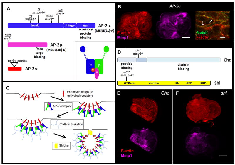
A: Coding regions of AP-2 complex subunits with allele locations and details and cartoon of AP-2 adaptor complex (inset). AP-2α is disrupted in the MENE(2L)-A complementation group, while AP-2μ is disrupted in the MENE(3R)-D complementation group. B: AP-2σ mutant L3 eye imaginal discs stained for F-actin (red), Mmp1 (magenta), and Notch (green) (left, middle, and right panels, respectively). C: Schematic of AP-2-dependent endocytosis and cell surface endocytic regulators. D: Chc and shi coding regions with location of mutant alleles. Chc (E) and shi (F) mutant L3 eye imaginal discs stained for F-actin (red) and Mmp1(magenta). Scale bars: 100μm (B, left and middle panels, E-F) and 10μm (B, right panel). Δ = deletion, fs = frameshift, * = stop codon.