
Figure 4. Endocytic Requirements for Delta/Notch Signaling in Signal-Receiving and Signal-Sending Cells.
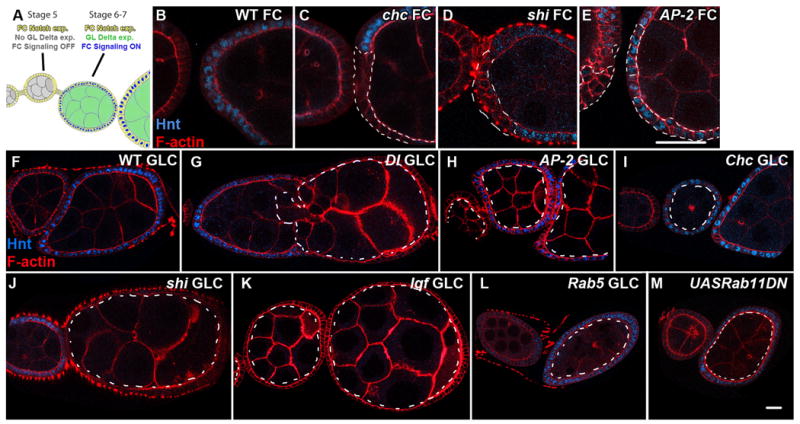
A: Schematic of Notch and Delta expression and signaling in the developing Drosophila ovary, adapted from [16]; Notch in follicle cells is activated at Stage 6/7 in response to Delta signaling from the germline. Staining for the Notch signaling target Hindsight (Hnt) (blue) in WT (B), Chc (C), shi (D), and AP-2α (E) follicle cell clones (FCs). Hnt staining is seen in AP-2α, but not Chc or shi mutant follicle cells. Hnt staining (blue) in WT follicle cells surrounding WT (F), Dl (G), AP-2α (H), Chc (I), shi(J), lqf (K), Rab5(L), and Rab11DN-expressing(M) germline clones (GLCs). Hnt is present in follicle cells surrounding AP-2α, Chc, Rab5, and Rab11-expressing, but not shi or lqf germline clones. All mutant clones are outlined by dashed lines. Scale bars: 30μm (B-E) and 20μm (F-M).