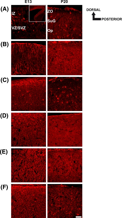
Fig. 5.
Spatiotemporal expression pattern of RPTPJ (a), RPTPκ (b), RPTPγ (c), RPTPε (d), RPTPRR (e) and RPTPσ (f) in the embryonic (E13) and mature (P20) mouse superior colliculus. Sagittal sections through the embryonic (E13) and late postnatal superior colliculus (P20) were labeled with appropriate RPTP-antibodies. Dorsal and posterior orientation is schematically presented. a At E13, a polyclonal antibody against RPTPJ detects single immunoreactive cells in the subventricular zone (SVZ) of the embryonic superior colliculus. In addition we found RPTPJ expressing cells in a decreasing anterior–posterior gradient (insert, Fig. 5a). Insert: lower magnification of a RPTPκ stained sagittal section of the E13 superior colliculus (scale bar = 200 μm). In the P20 superior colliculus, single cells, scattered throughout the stratum griseum superficiale (SuG) and stratum opticum (SO), were labeled. b The RPTPκ epitope is located on radial processes in the embryonic superior colliculus, whereas in the P20 superior colliculus a broad RPTPκ-immunoreactivity was found within all visual layers. (c, d and f) The strongest RPTPγ-, RPTPε- and RPTPσ-immunoreactivities were found in the dorsal part of the E13 superior colliculus (IZ intermediate zone). At P20, RPTPγ and RPTPσ immunoreactive cells were restricted to the stratum griseum superficiale (SuG) and stratum opticum (SO), whereas RPTPε immunoreactivity displays a more dispersed distribution pattern. e Immunolabeling using a polyclonal RPTPRR antibody reveals a weak and uniform staining pattern in the embryonic as well as in the P20 superior colliculus. IZ intermediate zone, VZ/SVZ ventricular zone/subventricular zone, ZO stratum zonale, SuG stratum griseum superficiale, Op stratum opticum. Scale bar 50 μm

