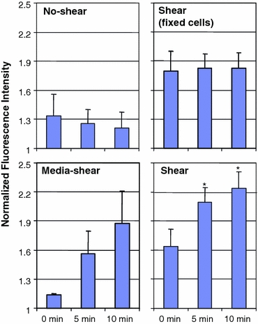
Figure 9.

Normalized GFP intensity in perinuclear compartment. The fluorescent intensities are normalized with respect to the average cell intensity at the same time point after background subtraction. Top left U937 cells not exposed to fluid shear do not show significant changes in GFP intensity level in the perinuclear compartment (n = 3). Top right Fixed U937 cells exposed to shear by pH-balanced Plasma-Lyte exhibit a constant GFP intensity in the perinuclear compartment for the duration of the experiment (n = 3). Bottom left U937 cells exposed to shear by culturing medium showed marked increase (but not statistically significant) of GFP intensity (n = 3). Bottom right U937 cells subjected to shear of pH-balanced Plasma-Lyte shows statistically significant (* p < 0.05) increase in GFP fluorescence compared to the 0 min time point (n = 8). Error bars = standard error
