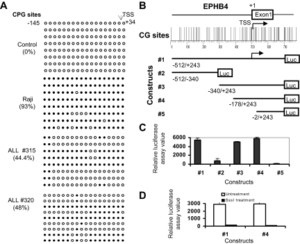
Figure 4.
Promoter hypermethylation silences expression of EPHB4. (A) Methylation status of EPHB4 gene was analyzed by bisulfite sequencing of the promoter region. Each row of circles represents the sequence of an individual clone; ○, unmethylated CpG sites; and ●, methylated CpG sites. (B) Diagram of the human EphB4 promoter region studied. CpG sites are indicated by short vertical bars. Arrows point to TSS. Below are serial deletion constructs of the EphB4 regulatory sequence. (C) Relative luciferase activity of different portions of the unmethylated EphB4 promoter constructs pGL3-EphB4 1, 2 3, 4, and 5 in 293T cells. Error bars indicate range. (D) Relative luciferase activities of unmethylated (□) and methylated (■) pGL3-EphB4 1 and 4 vectors in 293T cells. Promoter methylation of the EphB4 5′ region inhibits luciferase activity.

