
Figure 1. Drosophila neurons can span large distances in the brain.
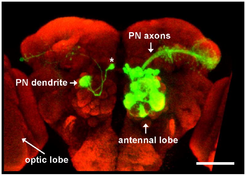
(a) On the right hemisphere of the brain a large number of PNs are labeled with GFP. On the left hemisphere a single PN is labeled revealing both its dendritic and axonal morphology (the cell body is indicated by the asterisk). Note the large distance the axon traverses. The optic lobes from both hemispheres of the brain are cropped in this image. The scale bar corresponds to approximately 50 μm. Modified with permission from Ref. 100.