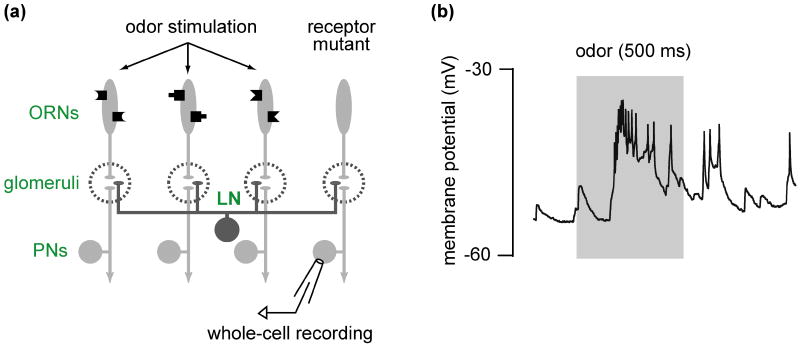
Figure 4. Functional dissection of synaptic inputs to a neuron.
(a) Experimental design for silencing one synaptic input in order to reveal others. The odor receptor gene expressed in one particular type of olfactory receptor neuron (ORN) is mutated, and therefore these neurons don't respond to odors. A whole-cell recording is made from a projection neuron directly postsynaptic to the mutant ORNs. An odor is used to stimulate the remaining functional ORN types. Local neurons (LN) are anatomically poised to mediate communication between the glomerular compartments.
(b) Example voltage trace from a PN postsynaptic to the silenced ORNs. Odor stimulation (gray bar) produces a depolarization leading to a train of spikes. Because this PN's direct presynaptic ORNs are silent, this excitation must derive from the activation of surrounding glomeruli, and therefore implicates local excitatory circuits in contributing to PN odor responses. (b) is modified with permission from Ref. 73.

