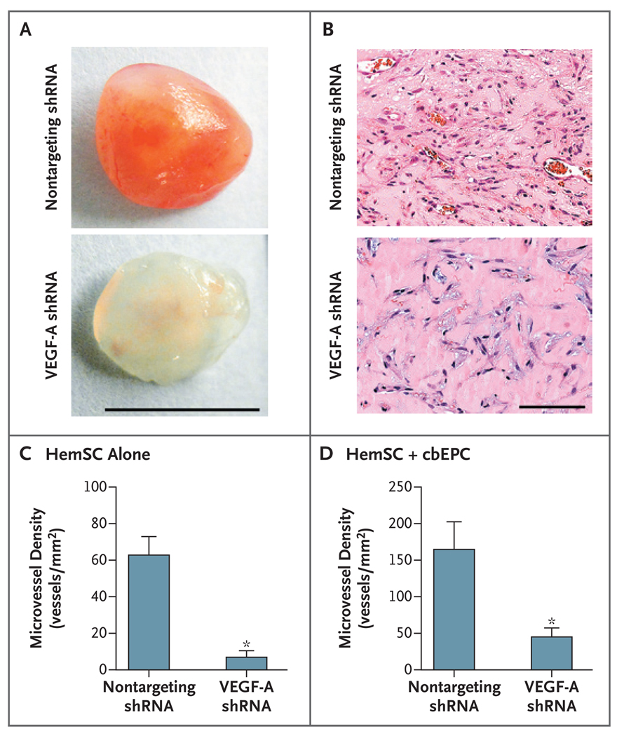
Figure 4. Inhibition of Vasculogenesis through Silencing of VEGF-A in Hemangioma-Derived Stem Cells.
Hemangioma-derived stem cells (HemSC) were stably infected with short hairpin RNA (shRNA) from vascular endothelial growth factor A (VEGF-A) or with a nontargeting shRNA (control). Hemangioma-derived stem cells (4×10⁶) that were infected with either VEGF-A shRNA or nontargeting shRNA were suspended in Matrigel and implanted subcutaneously in nude mice. Panel A shows representative Matrigel explants at day 7, with 7 to 10 samples in each subgroup. Panel B shows sections from representative explants from the VEGF-A shRNA group and the nontargeting shRNA group (hematoxylin and eosin). Panel C shows microvessel-density analysis of explants containing VEGF-A shRNA or nontargeting shRNA after 7 days in vivo. Panel D shows microvessel-density analysis of explants with the use of the two-cell model, in which either VEGF-A shRNA cells or nontargeting shRNA cells were combined with cord-blood endothelial progenitor cells (cbEPC), with eight samples in each subgroup. In Panels C and D, asterisks indicate that the comparison with the control group was significant. The T bars indicate standard errors. For both one-cell and two-cell models, the experiment was repeated twice with similar results.

