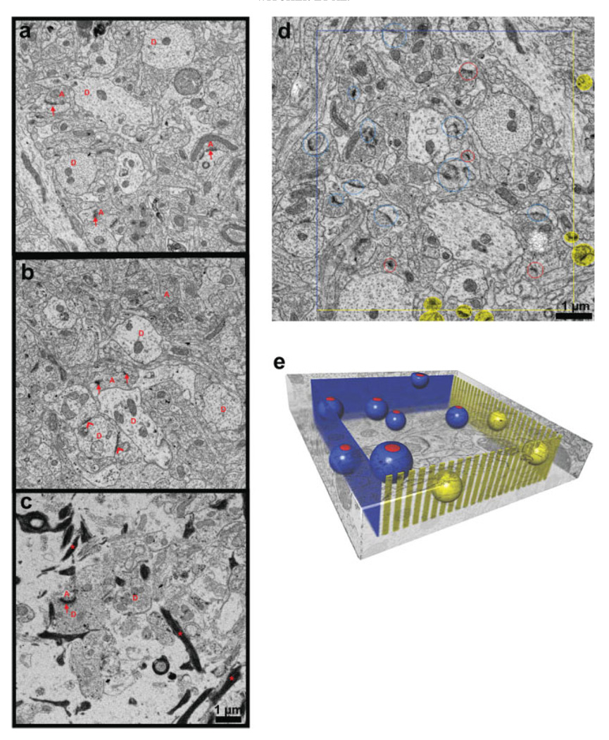
Fig. 2.
Ultrastructural images from each case and illustration of the unbiased method. Mild (a) and moderate (b) cases with compact neuropil and little extracellular space had dendrites [D] with intact cytoplasm, organelles, and well-ordered arrays of microtubules. Asymmetric (red arrows) and symmetric (red chevrons) synapses apposed to axonal boutons [red A]. (c) Severe case had neuropil with more extracellular space, fewer dendrites [D], axons [A], synapses (arrows), and astroglial processes filled with densely stained filaments (stars). Unbiased stereological analysis: (d) Central section of series from mild case, and (e) schematic of the unbiased analysis. Synapses counted if their PSDs were completely contained within sampling volume or intersected inclusion faces (blue lines in d, blue planes in e). Synapses excluded if they intersected exclusion faces (yellow lines d, yellow “picket fence” in e). Synapses intersecting bottom face were included and synapses intersecting top face were excluded (top and bottom faces not shown). Included synapses were encircled with red on the first section where the PSD appeared in the series and then circled with dark blue on adjacent serial sections to identify them and assure that none were double counted. Red and dark blue spheres illustrate included synapses and yellow spheres illustrate excluded synapses in e.

