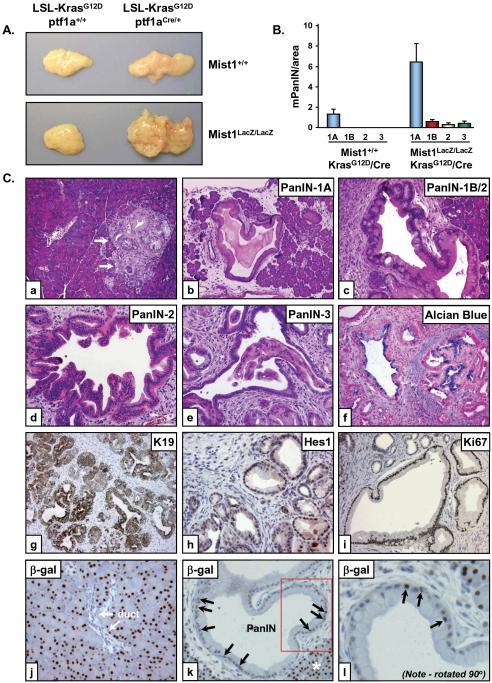
Figure 3. Mist1LacZ/LacZ/LSL-KrasG12D/+/ptf1aCre/+ pancreata display accelerated histological progression of mPanIN lesions at early stages.
(A) Gross anatomy of the Mist1LacZ/LacZ/LSL-KrasG12D/+/ptf1aCre/+ versus Mist1+/+/LSL-KrasG12D/+/ptf1aCre/+ pancreata at 6 weeks. (B) Quantitative analysis of mPanIN lesions in 6 week Mist1+/+/LSL-KrasG12D/+/ptf1aCre/+ and Mist1LacZ/LacZ/LSL-KrasG12D/+/ptf1aCre/+ samples (n=4). Note that high-grade mPanIN-1B, mPanIN-2 and mPanIN-3 are never observed in the Mist1+/+/LSL-KrasG12D/+/ptf1aCre/+ mice at this age. (C) High-grade mPanIN lesions rapidly develop in Mist1LacZ/LacZ/LSL-KrasG12D/+/ptf1aCre/+ pancreata. (a) 6-week Mist1+/+/LSL-KrasG12D/+/ptf1aCre/+ pancreas showing a rare focus of mPanIN-1A (arrows) (x100). (b-l) Representative images from Mist1LacZ/LacZ/LSL-KrasG12D/+/ptf1aCre/+ samples (6 week). mPanIN-1A lesions are broadly distributed (b, x100) with early appearance of higher grade mPanIN-1B (c, x200), mPanIN-2 (d, x200) and mPanIN-3 (e, x200) lesions. (f) Alcian blue staining reveals abundant mucin content in the mPanIN lesions. K19 (g), Hes1 (h) and Ki67 (i) are highly expressed in the Mist1LacZ/LacZ/LSL-KrasG12D/+/ptf1aCre/+ mPanIN lesions. (j) β-gal immunohistochemistry on Mist1LacZ/LacZ/LSL-KrasG12D/+ control samples revealing nuclear β-gal only in acinar cells. (k,l) β-gal positive nuclei are found within large PanINs from Mist1LacZ/LacZ/LSL-KrasG12D/+/ptf1aCre/+ samples (arrows). Boxed area in (k) is shown at higher magnification in (l). Asterisk in (k) denotes normal acinar tissue.

