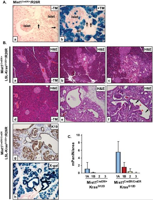
Figure 4. LSL-KrasG12D/+/Mist1Cre-ER/+ mice reveal that acinar cells generate mPanIN lesions.
(A) Mist1Cre-ER/+/R26R adult mice were given a single dose of corn oil (a) or tamoxifen (TM) (b) for three consecutive days and then pancreata were harvested and processed for β-gal detection 2 weeks later. Acinar cells and rare islet cells are β-gal+ whereas all duct cells (arrows) remain β-gal negative (x400). (B) Adult 6 week LSL-KrasG12D/+/Mist1Cre-ER/+/R26R (a-c) and LSL-KrasG12D/+/Mist1Cre-ER/Cre-ER/R26R (d-f) mice were administered corn oil (a,d, x200) or tamoxifen (b,c, x100; e,f, x200) and pancreata were analyzed at 3 months of age. LSL-KrasG12D/+/Mist1Cre-ER/Cre-ER/R26R pancreata develop advanced mPanINs which express high levels of K19 (g, x200). (h) β-gal+ mPanINs (arrows) confirm the acinar cell origin of these lesions (x200). Arrows in (f) indicate mPanIN extensions (red) and fusion with normal ductal epithelial (black). (C) Quantitative analysis of mPanIN lesions in 6 week LSL-KrasG12D/+/Mist1Cre-ER/+ and LSL-KrasG12D/+/Mist1Cre-ER/Cre-ER mice treated with tamoxifen (n=4). Note the large increase in all mPanIN grades in the LSL-KrasG12D/+/Mist1Cre-ER/Cre-ER (Mist1KO) mice.

