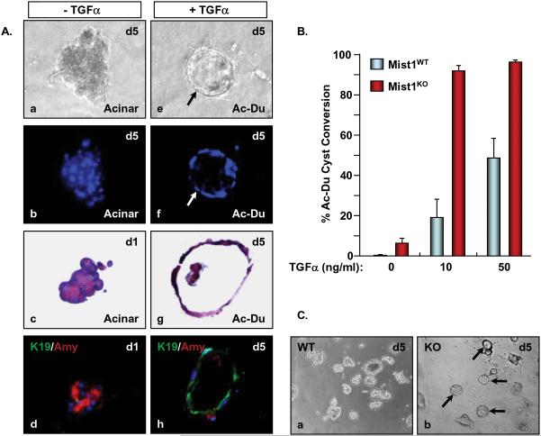
Figure 6. Mist1LacZ/LacZ acinar cells rapidly convert to ductal cysts in 3D collagen cultures.
(A) Acinar cells were isolated from Mist1+/+ pancreata and cultured in collagen gels in the presence or absence of TGFα. Cells in control medium without TGFα (a-d, x800) maintain a normal amylase positive acinar cell phenotype while cells supplied TGFα (e-h, x800) convert to K19 positive ductal cysts (arrow) (b,f - Dapi fluorescence; c.g - H&E sections; d,h - K19 and amylase co-immunofluorescence). (B) Mist1LacZ/LacZ acinar cells exhibit a propensity to convert to ductal cysts, even in the absence of TGFα. (C) Nearly all Mist1LacZ/LacZ acinar cells convert to ductal cysts after 5 days in 10 ng/ml TGFα whereas Mist1+/+ cells remain as acinar cell clusters (x200).

