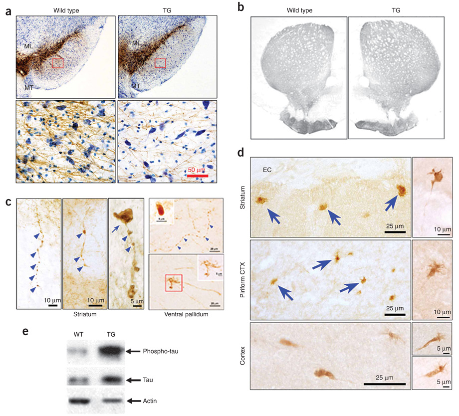
Figure 3.
Morphologic abnormalities of mesencephalic dopamine neurons and their axons in LRRK2R1441G BAC transgenic mice. (a) Tyrosine hydroxylase immunostaining of ventral mesencephalon. ML, medial lemniscus; MT, medial terminal nucleus. Rectangles in the upper panels are shown at a higher magnification in the lower panels. LRRK2R1441G BAC transgenic mice showed a loss of tyrosine hydroxylase–positive dendrites in substantia nigra pars reticulata and a reduction of tyrosine hydroxylase neuron size in SNpc. (b) Normal optical density of tyrosine hydroxylase immunostaining of the whole striatum and dorsal-lateral quadrant. (c) In tyrosine hydroxylase–positive axons in the striatum and the ventral pallidum of LRRK2R1441G BAC transgenic mice, we observed discrete foci of thickening with a beaded appearance (blue arrowheads) and fragmentation, large tyrosine hydroxylase–positive spheroid-like structures (blue arrow in the left panel and the insert in the upper right panel), and dystrophic neurites and enlarged axonal endings (lower right). The dystrophic neurites were found in all of the five LRRK2R1441G mice (2.8 ± 0.6, s.e.m.) but were rarely seen in two of the seven nontransgenic littermates (0.4 ± 0.3; P = 0.003, t test). Dystrophic neurites were not found in the two WT-OX mice. Thus, expressed as number of neurites per area of the ventral pallidum (mm2), dystrophic neurites were 8.5-fold more prevalent among the transgenic mice than the controls. (d) In the dorsal striatum and piriform cortex, immunohistochemistry with AT8 revealed abnormal axon terminal enlargements and dystrophic neurites (arrows, top two left panels). EC, external capsule. AT8-positive structures are shown at a higher magnification in the right panels. A total of 42 AT8-positive dystrophic neurites were identified in the two LRRK2R1441G mice that we examined. A single example was identified under blind conditions in one of the four nontransgenic mice, and none were found in the two WT-OX mice. Expressed as the number of neurites per forebrain coronal hemisection, 21 AT8-positive neurites were observed among the LRRK2R1441G transgenics and only 0.17 among the control brains, a 124-fold difference. (e) Phosphorylated tau was markedly increased in the LRRK2R1441G BAC transgenic mouse brains. Full-size western blots are presented in Supplementary Figure 7 online.

