Fig. 1.
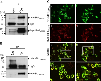
Coimmunoprecipitation and colocalization of Slo1VEDEC and Slo1QEERL in HEK293T cells. Lysates of HEK293T cells heterologously expressing Myc-tagged Slo1VEDEC and HA-tagged Slo1QEERL were immunoprecipitated with either mouse anti-Myc (A) or anti-HA antibodies (B). Normal mouse IgG served as a negative control. The immunoprecipitated Slo1 proteins were detected with anti-HA or anti-Myc as indicated. C, colocalization of HA-tagged Slo1VEDEC and Myc-tagged Slo1QEERL in HEK293T cells visualized by confocal microscopy. Slo1VEDEC was detected with anti-HA (green, a and b), and Slo1QEERL was detected with anti-Myc (red, c and d). Merged signals of Slo1VEDEC and Slo1QEERL are shown in e to h. Boxed regions in e and f are magnified in g and h, respectively. Colocalization in merged images appear as a yellow signal.

