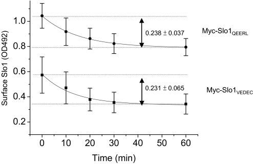
Fig. 9.
Time course of Slo1VEDEC and Slo1QEERL removal from the cell surface. Endocytosis assays were carried out by using HEK293T cells heterologously expressing either Slo1VEDEC or Slo1QEERL. The expressed channels bear an Myc tag at the extracellular NH2 terminus, which allowed surface Slo1 on the surface of intact cells to be labeled by anti-Myc at 4°C. Cells were then placed at 37°C for various times to allow trafficking to resume, at which time they were fixed. The amounts of anti-Myc remaining on the cell surface were determined by HRP-conjugated anti-mouse with colorimetric assays at OD492. Data show the time course of OD492 (Mean ± S.E.M.) from three different experiments. ■, Slo1VEDEC; ●, Slo1QEERL. Data are fitted with single-exponential decay functions with time constants of 15.1 ± 0.9 (for Slo1QEERL) and 14.4 ± 3.4 min (for Slo1VEDEC).

