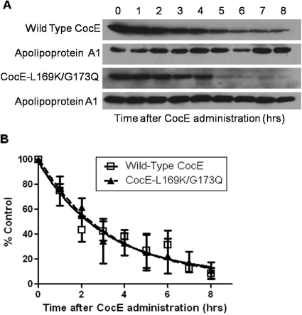
Fig. 5.
In vivo CocE plasma half-life. A, representative Western blots of CocE from mouse serum over time. CocE (L169K/G173Q or wt) was administered to mice intravenously via the lateral tail vein. Blood samples were taken at the times indicated by submandibular sampling. Serum was collected and 20 μg of total serum protein was run on a 10% SDS polyacrylamide gel. Blotting was performed with rabbit anti-CocE antibody and rabbit anti-Apolipoprotein A1 antibody. Wild-type and CocE-L169K/G173Q were both tested in three independent groups of animals followed by serum analysis. B, quantification of wt and CocE-L169K/G173Q Western blot densities analyzed with Image J software. All time points are adjusted as a fraction of the apoA1 loading control. Fit to a one-phase exponential decay model, the half-life of CocE-L169K/G173Q was determined to be 2.3 h after administration (wt = 2.2 h). two-way ANOVA, p = 0.92.

