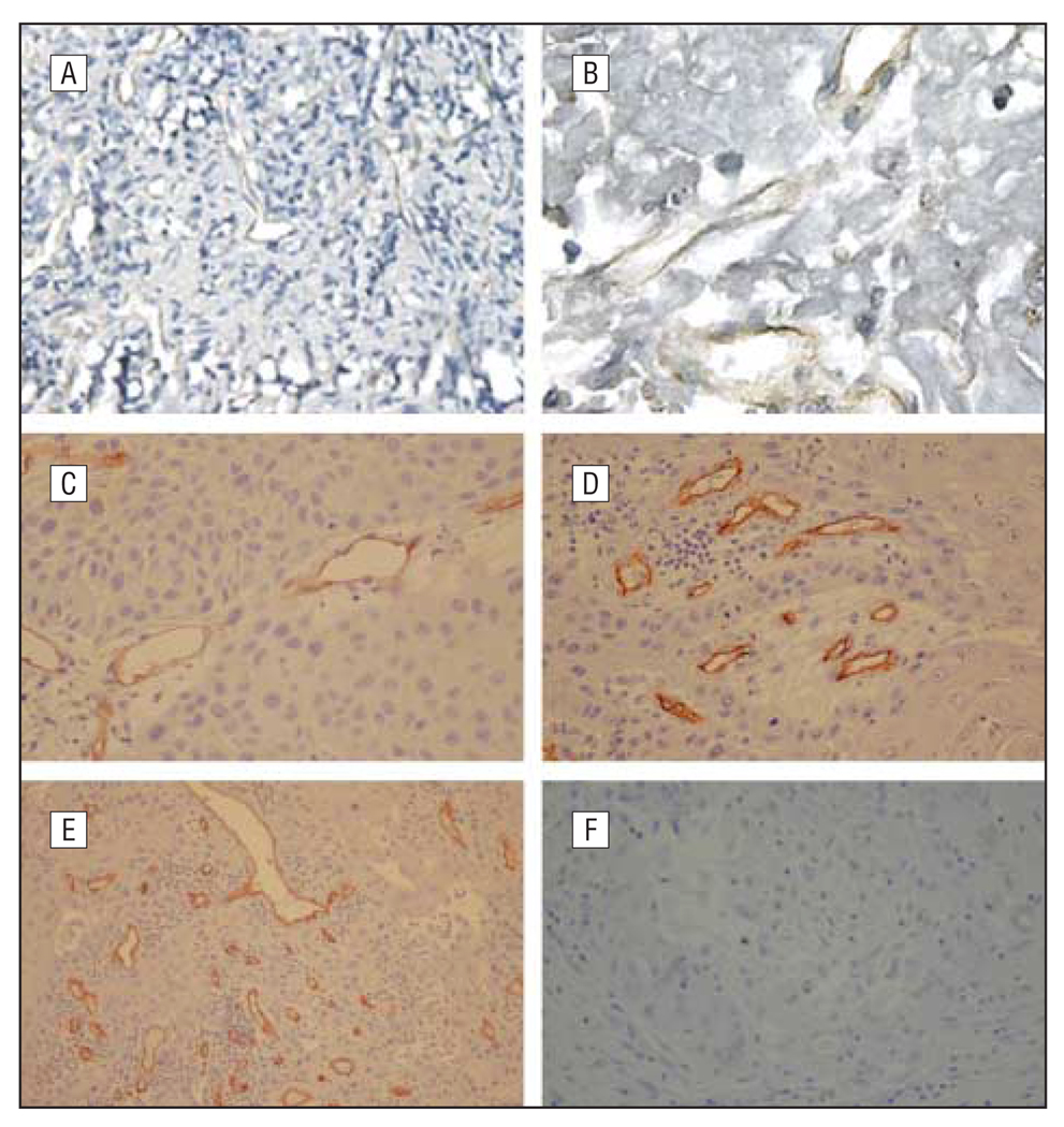
Figure 3.
Identification of microvessels. Identification of microvessels in head and neck squamous cell carcinoma (HNSCC) specimens with endothelial cell markers. Specific von Willebrand factor (vWF) and CD34-specific antibodies were used for staining HNSCC specimens. Microvessels were observed in the HNSCC specimens at various densities. A and B, Anti-vWF antibody weakly stained the microvessel endothelial cells (original magnifications ×100 [A] and ×400 [B]). C–E, Anti-CD34 clearly stained the endothelial cells in the HNSCC specimens (original magnifications ×200 [C], ×200 [D], and ×100 [E]). F, Nonspecific IgG did not stain (original magnification ×100).

