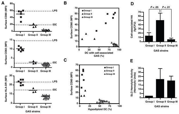
Figure 1.
GAS strains vary in their capacity to induce DC maturation or apoptosis. Human monocyte-derived DC were exposed to live GAS for two h at a GAS to DC ratio of 1:1. Twenty h later, surface expression of CD80, CD83, and HLA-DR was quantified by flow cytometry (A). Values for control untreated DC, and LPS-stimulated DC are shown as dashed lines. Following staining of surface CD80, DC were fixed, permeabilized, and stained with a GAS-specific antiserum to assess binding and internalization of GAS (B), or with propidium iodide to determine percentage of hypodiploid nuclei (apoptotic cells) (C). Each symbol represents the mean of results for three independent donors. 10,000 cells were examined in each measurement. GAS clinical isolates were classified into three groups according to DC responses to each strain: group I, mature DC (filled squares); group II, immature DC (filled triangles); group III, immature apoptotic DC (open triangles). To further characterize the phenotype of GAS strains, bacterial cell-associated HA (D) and SLO-hemolytic activity (E) of the GAS strains were determined. Bars represent mean values±SD of at least three independent experiments for GAS clinical isolates included in each group.

