Figure 4.
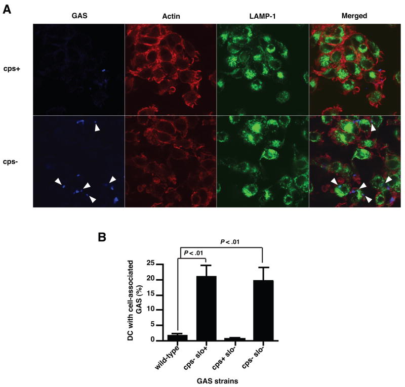
The HA capsule prevents binding an internalization of GAS by DC. A, Confocal microscopy analysis of GAS phagocytosis. Human DC pulsed with live GAS for 2 h were allowed to attach to poly-L-lysine coated cover slips, fixed, and permeabilized prior to staining with GAS-specific antiserum (blue). The lysosomal antigen LAMP-1 (green) and the actin cytoskeleton (red) were also visualized. Arrowheads show intracellular GAS. B, FACS analysis of GAS phagocytosis by DC. GAS-pulsed human DC were harvested 2 h post-inoculation, stained with GAS-specific antiserum, and analyzed by flow cytometry. Percentage of DC positive for GAS staining was compared to untreated DC. Means±SD of results from four independent donors are shown.

