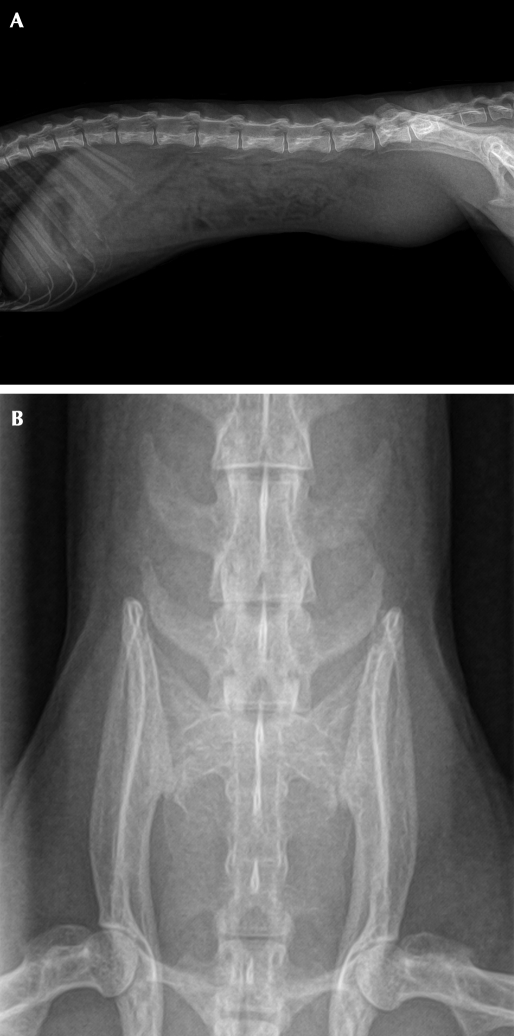
Figure 2.
(A) Right lateral radiographic view of squirrel monkey. (B) Ventrodorsal view of the caudal abdomen of squirrel monkey. In both views, the caudal abdominal mass appears as a round opacity with ill-defined borders; it is superimposed over the sixth and seventh lumbar vertebrae in the ventrodorsal view. Whether surrounding structures within the caudal abdomen were involved could not be determined by imaging.

