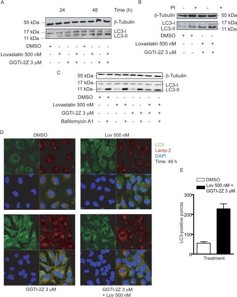
Fig. 7.
Induction of autophagy in STS-26T cells by GGTI-2Z/lovastatin treatment. STS-26T cells were treated with the indicated concentrations of GGTI-2Z and lovastatin alone or in combination. Results are representative of three independent experiments. A, treatments were for 24 or 48 h as shown. Whole-cell lysates were then probed for LC3 and β-tubulin. B, cells were subject to 2-h pretreatment with protease inhibitors, 10 μM pepstatin A, and 10 μM E64D as indicated. Whole-cell lysates were then probed for LC3 and β-tubulin. C, cells were subject to 48 h of drug treatment, with addition of 50 nM bafilomycin A1 for the last 2 h of the incubation as indicated. Whole-cell lysates were then probed for LC3 and β-tubulin. D, STS-26T cells were treated as indicated for 48 h followed by methanol fixation. Cells were then stained for LC3 and LAMP-2. Nuclei were stained with 4′,6-diamidino-2-phenylindole (DAPI), and cells were visualized under a LSM-510 microscope at 40× magnification. E, quantitative analysis of LC3-positive puncta treated with either DMSO or the drug combination for 48 h.

