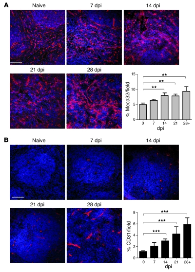
Figure 2. Increased expression of Meca32 and CD31 in L. donovani–infected spleens.
(A and B) Expression of Meca32 (A) and CD31 (B) was determined (red) by immunostaining of frozen spleen sections from naive mice and mice at 7, 14, 21, and 28 dpi. Sections were counterstained with the nuclear dye DAPI (blue). Scale bars: 100 μm. Quantification of the area covered by Meca32+ and CD31+ endothelial cells in whole spleen was determined using computer-assisted morphometric analysis. A significant increase in the vascular area was observed in tissues beginning at 14 dpi compared with naive animals. Data are mean ± SEM of at least 3 independent experiments (n = 20 per time point). **P = 0.01, ***P < 0.0001, ANOVA.

