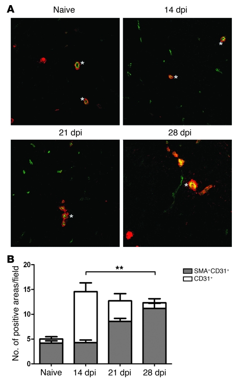
Figure 3. Vessel maturation during L. donovani–induced vascular remodeling.
(A) Sections of frozen spleens taken during the course of L. donovani infection were stained with antibodies to CD31 and α-SMA. Fluorescent images highlight costaining of CD31+ vessels (green) and α-SMA (red) on splenic white pulp vessels in naive mice and mice at 14, 21, and 28 dpi. Asterisks denote central arterioles. Note the increased expression of α-SMA on the CD31+ vessels in the chronic stages of infection. Original magnification, ×200. (B) Quantification of CD31/SMA association during the course of L. donovani infection. The number of areas positive for CD31 was counted per field of view, and the number with intimately associated SMA staining was then determined. The combined data highlight the differences in the ratio of CD31+ to SMA+CD31+ areas. At least 3 fields of view were counted per spleen section. Data are mean ± SEM of at least 2 independent experiments (n = 3 per time point). **P = 0.0012.

