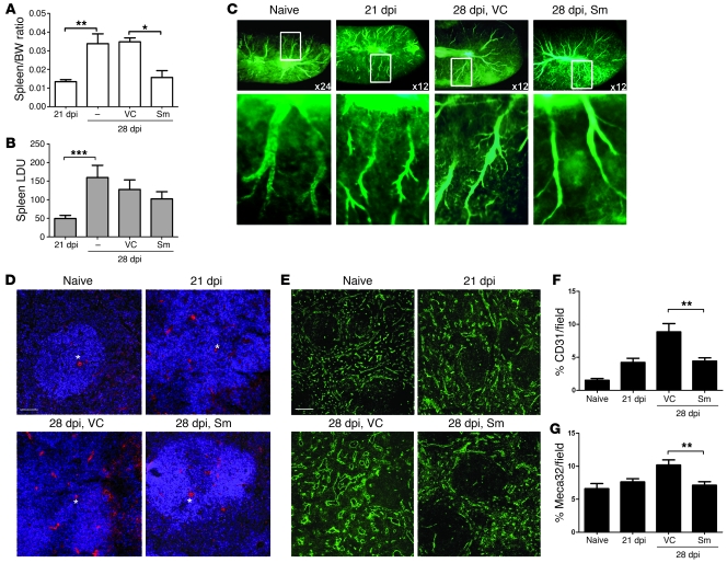
Figure 4. RTKI treatment inhibits pathogen-induced splenic vascular remodeling.
(A) Spleen/body weight ratios in 21- and 28-dpi L. donovani–infected mice, measured before and after Sm treatment compared with vehicle control (VC) or no treatment. The significant increase in spleen weights and spleen/body weight ratios observed at 28 dpi compared with 21 dpi was inhibited by Sm treatment compared with vehicle control treatment. *P = 0.03, **P = 0.008. (B) Splenic parasite burdens were not altered by Sm treatment. ***P = 0.006. (C) Changes in splenic vasculature after Sm treatment, assessed by FITC-dextran angiography. Boxed regions are shown at higher magnification below, showing details of vascular branching. Original magnification, ×24 (naive, top); ×120 (naive, bottom); ×12 (infected, top); ×60 (infected, bottom). (D) Effect of Sm treatment on expression of CD31 (red) in L. donovani–infected spleens. Sections were counterstained with DAPI (blue). Scale bars: 100 μm. (E) Effect of Sm and vehicle control treatment on the expression of Meca32 (green) in L. donovani–infected compared with naive mouse spleens. Scale bars: 100 μm. (F and G) Area covered by CD31+ (F) and Meca32+ (G) endothelial cells in whole spleen following Sm treatment, determined using computer-assisted morphometric analysis. A significant decrease in the CD31+ (**P = 0.005) and Meca32+ (**P = 0.004) vascular areas was observed in tissues from Sm-treated mice compared with vehicle control–treated mice. Data are mean ± SEM of at least 3 independent experiments (n = 5 per time point).

