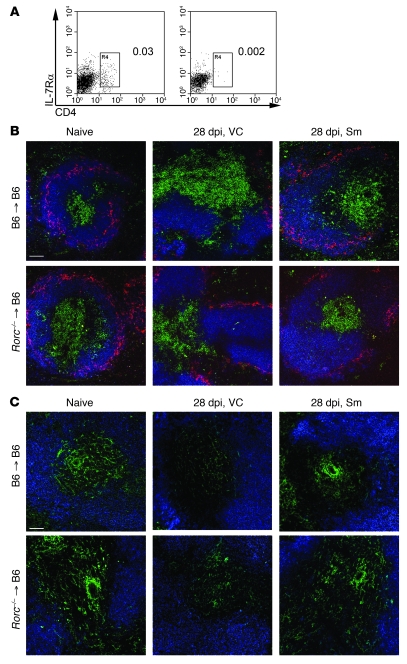
Figure 6. Restoration of splenic architecture after RTKI treatment is not dependent on LTi cells.
(A) Flow cytometry analysis of LTi cells in the spleen of B6.Rorc–/–→B6.CD45.1 chimeric mice (LTi insufficient; right) and B6.CD45.1→B6.CD45.1 chimeric mice (LTi sufficient; left). Plots are gated on CD11c–B220–CD3–CD45+ cells; numbers denote percent LTi cells (IL-7Rα+CD4+) in spleen, as outlined by the boxed regions. Data are from 1 representative experiment (n = 3 per group). (B) Immunofluorescent staining of frozen spleen sections (10 μm) from naive and L. donovani–infected B6.Rorc–/–→B6.CD45.1 and B6→B6.CD45.1 chimeras after treatment with Sm or vehicle control, highlighting localization of B cells (B220, blue), T cells (CD3, green), and MMM (CD169, red). (C) Splenic T cell zones depicting FRC (gp38, green) and B cells (B220, blue). Scale bar: 100 μm.

