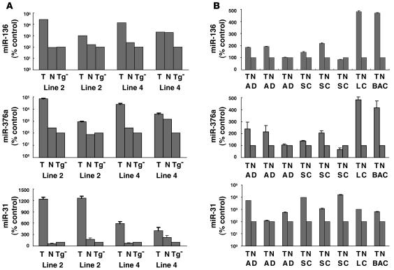
Figure 3. Validation of miR-136, miR-376a, and miR-31 expression profiles by real-time RT-PCR assays performed on RNA isolated from the indicated murine cyclin E–transgenic lines and from paired human normal-malignant lung tissues.
(A) Real-time RT-PCR assays for miR-136, miR-376a, and miR-31 were performed. T, malignant tumor; N, adjacent normal murine lung; and Tg–, murine nontransgenic FVB normal lung tissue. Results were normalized to expression levels detected in FVB murine Tg– lung tissues. (B) Real-time RT-PCR assays for miR-136, miR-376a, and miR-31 were independently performed on paired human normal-malignant lung tissues. AD, adenocarcinoma; SC, squamous cell carcinoma; LC, large cell carcinoma; BAC, bronchoalveolar carcinoma. Results were normalized to expression levels measured in normal human lung. Error bars indicate SD.

