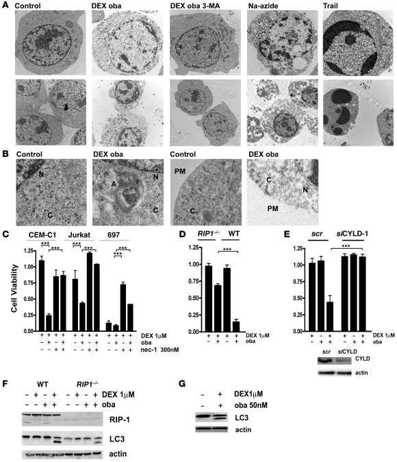
Figure 7. RIP-1 kinase activity is essential for obatoclax-mediated GC sensitization.
(A) Electron microscopy images reveal features of necroptotic cell death after treatment for 72 hours with obatoclax and dexamethasone. No condensed chromatin was detectable, while Trail treatment induces condensed apoptotic nuclei (top panel) (original magnification, ×7,100). Cells treated with obatoclax and dexamethasone exhibited disintegrated plasma membranes, which was recapitulated by Na-azide. Trail treatment leaved membranes intact (bottom panel) (original magnification, ×5,400). (B) A more detailed view of the same experiment. An autophagosome formation with characteristic double membrane structures was detected in the cytoplasm. The plasma membrane was disrupted in cells treated with obatoclax and dexamethasone. N, nucleus; C, cytoplasm; A, autophagosome; PM, plasma membrane (original magnification, ×15,000). (C) In steroid-resistant (CEM-C1 and Jurkat) and steroid-sensitive (697) cells treated for 72 hours with dexamethasone (1 μM) and obatoclax (10% IC50), with or without the necroptosis inhibitor nec-1 (300 nM), nec-1 restored steroid resistance as assessed by the MTT assay. (D) Jurkat RIP1–/– cells were less sensitive to the double treatment for 72 hours with dexamethasone (1 mM) and obatoclax (10% IC50) compared with WT Jurkat cells. Cell viability was assessed by the MTT assay. (E) Downregulation of CYLD rescued Jurkat cells from cell death induced by combination treatment for 72 hours with dexamethasone (1 μM) and obatoclax (10% IC50). Efficiency of downregulation was assessed by Western blot analysis after 48 hours. (F) LC3-II generation occurred in Jurkat RIP1–/– cells and in WT cells after 8 hours of treatment with dexamethasone and obatoclax. (G) Treatment with nec-1 did not inhibit LC3-II generation induced by treatment with obatoclax and dexamethasone in Jurkat cells. ***P < 0.05.

