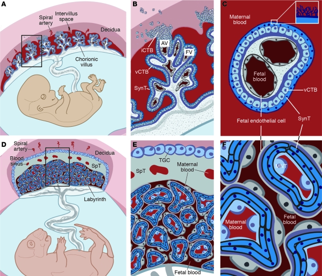
Figure 1. Comparative anatomy of human and mouse placentas.
Placentation is categorized by the relationship between trophoblasts and the maternal cells/tissues with which they come into contact. In the hemochorial placentas of humans (A–C) and mice (D–F), the maternal vessels are invaded and colonized by invasive trophoblasts (not shown). (A) Consequently, in humans, decidual spiral arterioles perfuse the chorionic villi that line the intervillous space (adapted from PLoS Pathogens; ref. 158). (B) In floating villi (FV), a continuous layer of multinucleated SynT interfaces with maternal blood. Beneath lies a progenitor population of mononuclear vCTB. At the uterine wall, iCTBs differentiate along the invasive pathway to form anchoring villi (AV). A subset of iCTBs breaches spiral arterioles and differentiates into an endovascular subtype that replaces the resident maternal endothelium (not shown). (C) The cross-sectional anatomy of a floating villus shows that the apical surfaces of SynTs are covered with branched microvilli that maximize their surface areas for gas and nutrient/waste exchange. The blood vessels that ramify through the villous stroma carry embryonic/fetal blood. (D) In mice, maternal blood from decidual spiral arterioles flows through blood sinuses in the SpT layer to reach the labyrinth. (E) TGCs, like iCTBs, anchor the placenta to the uterus and invade the spiral arterioles (not shown). (F) In mice, maternal blood is in direct contact with a layer of mononuclear trophoblasts (MNTs, also known as S-TGCs) that is surrounded by a bilayer of SynTs, which are in close proximity to fetal capillaries.

