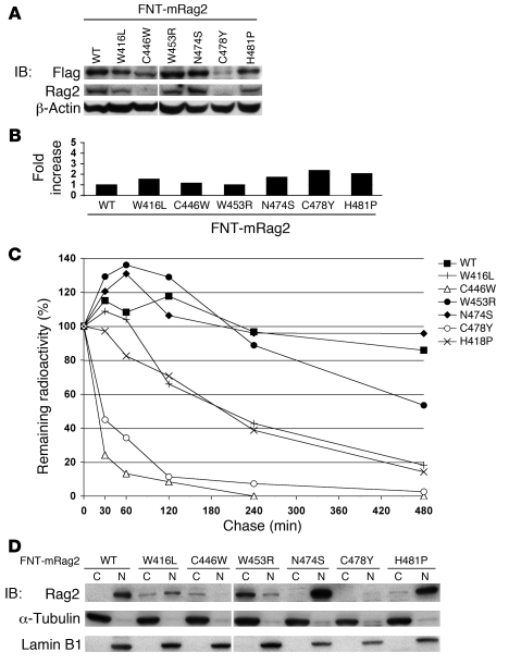
Figure 2. T-B-SCID/OS Rag2PHD mutations affect Rag2 stability and cellular localization in pro-B cells.
A mouse Rag2–/– pro-B cell line was retrovirally transduced to express FNT-tagged full-length wild-type Rag2 or T-B-SCID/OS mutants. (A) Western blot analysis was performed on whole cell extracts from retrovirally transduced pro-B cell lines for Flag, Rag2, and β-actin. Samples were loaded on the same gel, and the length of exposure was identical. The vertical line between FNT–mouse Rag2–C446W and –W453R mutants indicates a sample that was excluded from our study. (B) Total mRNA was isolated from retrovirally transduced pro-B cell lines, and Rag2 transcription levels were determined by Q-PCR. The average of 2 independent experiments are graphed as fold increase compared with mRNA expression of FNT–mouse Rag2. (C) Retrovirally transduced pro-B cell lines were pulse labeled with [35S] methionine/cysteine to determine degradation of wild-type Rag2 and T-B-SCID/OS mutants. Radiolabeled proteins were IP at various times after pulse with anti-Rag2 antibody, fractionated by SDS PAGE, and quantified on a PhosphorImager. Data were normalized to the radioactivity levels at the end of pulse. The graph presents the average from 2 independent experiments. (D) Cellular localization of Rag2 in retrovirally transduced pro-B cell lines was determined by Western blotting analysis of fractionated cytoplasm (C) and nuclear (N) extracts (top). Purity and loading of the fractions were analyzed by Western blotting for α-tubulin (middle) and Lamin B1 (bottom). The vertical line between FNT–mouse Rag2–C446W and –W453R is as in A.

