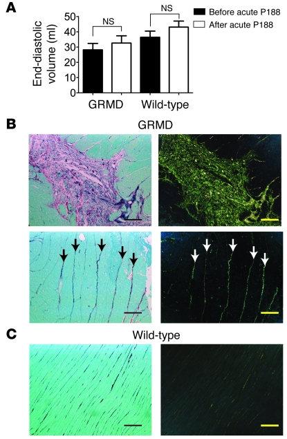
Figure 4. Preexisting fibrotic lesions and ventricular volume in GRMD dogs.
(A) Assessment of end-diastolic volume before and after acute infusion of P188 in GRMD and wild-type canines. Values are shown as mean ± SEM. (B) Representative Sirius red–stained histopathological sections from untreated adult GRMD animals. In brightfield images (left), collagen appears red; in polarized light, Sirius red–stained collagen exhibits birefringence, allowing it to be visualized as green-yellow light (right). Upper images show a large lesion with extensive fibrosis and degenerating myocytes. Lower images show strands of collagen that extend through areas of relatively normal myocardium (arrows). (C) Identical stains of wild-type myocardium. Scale bars: 100 μm.

