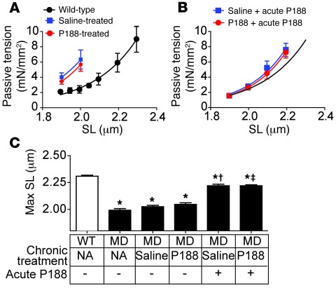
Figure 8. In vitro passive tension-extension relationships in intact GRMD cardiac myocytes after chronic infusion.
After the 8-week infusion, myocytes were isolated from P188- (red) and saline-infused (blue) GRMD and wild-type dogs. Passive tension-extension relationships of dystrophic cardiomyocytes in the absence (A) and presence (B) of acute application of 150 μM P188 are shown. The black curve in B is derived from wild-type myocyte data in A and is included as a reference. SL, sarcomere length. (C) Comparisons of maximum stable sarcomere length (Max SL) between the groups. *P < 0.05 versus wild-type; †P < 0.05 versus chronic saline GRMD group; ‡P < 0.05 versus chronic P188 GRMD group. Values are shown as mean ± SEM. All points were derived from 5–7 myocytes isolated from 4–5 dogs. All data were analyzed with 1-way ANOVA and Bonferroni’s multiple comparisons post-hoc test. MD, GRMD.

