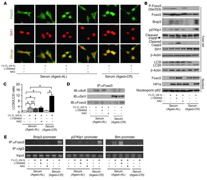
Figure 7. Involvement of PI3K and Sirt1 in Foxo3-mediated cell adaptation to hypoxia.
(A) Immunostaining showing localization of Foxo3 and Sirt1 at late phase of hypoxia (1% O2, 24 hours) under the indicated conditions. Original magnification, ×400. (B) Phosphorylation and expression level of Foxo3; expression of Bnip3, p27Kip1, cleaved PARP, cleaved caspase 3, and Sirt1; formation of LC3II; and nuclear expression levels of Foxo3 and Hif1a at late phase of hypoxia. To detect LC3I and LC3II bands, cells were preincubated with lysosomal inhibitor (E64d and pepstatin A). (C) Quantitative analysis of the ratio of LC3II to LC3I (n = 4). (D) Acetylation of Foxo3 and interaction between Sirt1 and Foxo3 at late phase of hypoxia. (E) ChIP analysis to determine Foxo3 binding to the promoters of Bnip3, p27Kip1, and Bim at late phase of hypoxia. LY294002 was used as a PI3K inhibitor at 20 μM. NAC was used as an antioxidant at 20 mM. Data are mean ± SEM. *P < 0.05.

