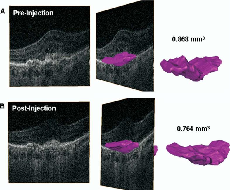
Figure 3.
A, Three-dimensional reconstruction of hsUHR-OCT data from before treatment. The pink object represents a volumetric reconstruction of the CNV lesion (both classic and occult components). Volume calculation shows the CNV to measure 0.868 mm3. B, Three-dimensional reconstruction of hsUHR-OCT data from 1 month after treatment. The pink object represents a volumetric reconstruction of the CNV lesion (both classic and occult components). Volume calculation shows the CNV to measure 0.764 mm3. CNV = choroidal neovascularization; hsUHR-OCT = high-speed ultrahigh resolution optical coherence tomography.

