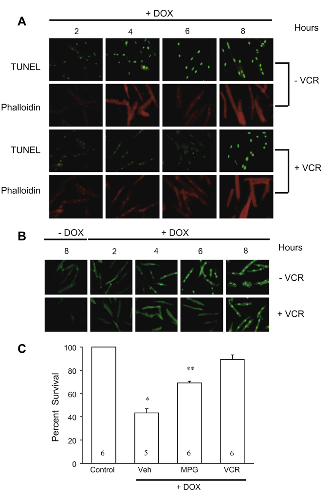
Fig. 4.
(A) Results of TUNEL staining to assess changes in the magnitude of apoptosis with doxorubicin treatment alone and co-treatment with vincristine. Co-treatment with vincristine delayed the onset of apoptosis. Phalloidin, which stains actin fibers, was used to confirm that the cells studied were rod-shaped myocytes. DOX, doxorubicin; VCR, vincristine. (B) Effects of doxorubicin treatment alone and with vincristine co-treatment on PARP activity monitored by assessment of poly (ADP)ribose (PAR) generation. The appearance of PAR was delayed during vincristine co-treatment. DOX, doxorubicin; VCR, vincristine. (C) Vincristine compared to the antioxidant N-(2-mercaptoproprionyl)-glycine) (MPG). Compared to 0.1 mmol/L MPG, the magnitude of cell survival (trypan blue exclusion assay) with vincristine (VCR, 15 µg/ml) was greater. Numbers in bars represent the number of experiments performed for each of the treatments depicted. Veh, vehicle; *p < 0.01 vs. all other bars; **p < 0.01 vs. Veh and VCR + DOX.

