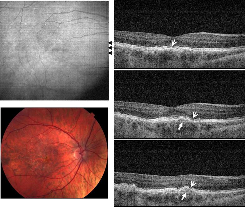
Fig. 2.
Image data from the right eye of a 58 year old female subject diagnosed with dry AMD in both eyes (View 2). Her right eye had a visual acuity of 20/25. The OCT cross-sectional images show elevation of the RPE due to drusen and changes in the photoreceptor inner and outer segment (IS/OS) boundary (thin arrows). Bruch's membrane appears under the area with large drusen deposits (solid arrows), but is not visible in the normal areas of the retina.

