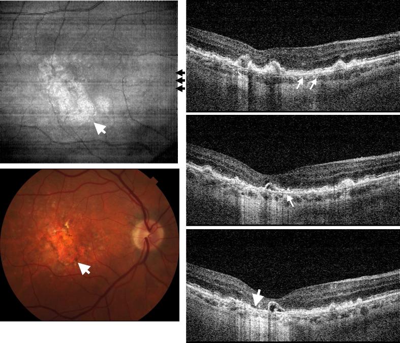
Fig. 5.
Image data from the right eye of a 67 year old male with dry AMD (View 5). Drusen deposit creates identifiable RPE and photoreceptor irregularities. The hyperscattering signal from the choroid beneath the lesion implies RPE atrophy. The IS/OS junction and RPE were no longer separable for UHR-OCT frames from the lesion area. Instead, the Bruch's membrane appeared under the RPE with a smooth contour.

