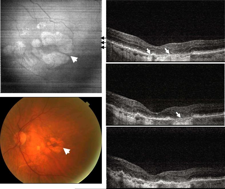
Fig. 6.
Image data from the left eye of an 84 year old female subject diagnosed with geographic atrophy and a visual acuity of 20/160 (View 6). Hyper-scattering appears on OCT images beneath the disrupted photoreceptor /RPE layers where Bruch's membrane can be easily identified. Both color photo and en face image of OCT demonstrate similar highly scattering area of RPE atrophy.

