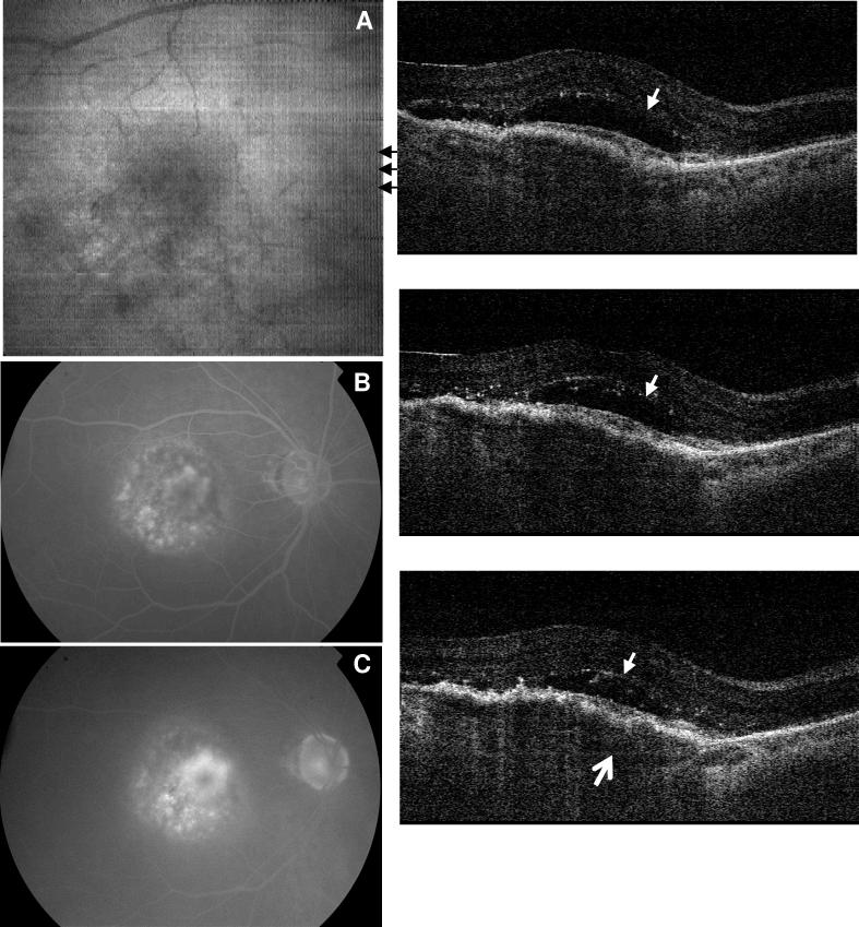
Fig. 7.
Image data from the right eye of a 77 year old female subject with wet AMD (View 7). Visual acuity was counting fingers at four feet. Fluorescein angiograms at 67 seconds (B) and 5 minutes and 8 seconds (C) indicate the development of choroidal neovascularization. A large volume of intraretinal fluid (small arrows) was identified from the OCT frames. The fluid developed over a large PED where the bottom arrow points the detached Bruch's membrane.

