
Figure 2.
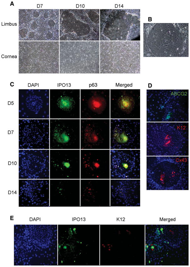
IPO13 expression in corneal epithelial clonal culture compared with other proposal markers. (A): Limbal and corneal epithelial clonal culture at different stages show that the number and size of clones in limbal epithelial clonal culture increased as the culture time prolonged. However, no clones were formed from the beginning to the end in corneal epithelial clonal cultures. (B): The cell size in the center of clone was much larger than that in the margin. (C): Immunofluorescent double staining for IPO13 and P63 in different stages clones showed that both IPO13 and P63 were expressed by almost all of the cells in early-stage clones. IPO13 was expressed in nuclei in the margin but in cytoplasm in the center of early-stage clones (D5, D7). IPO13-positive expression decreased as the clonal culture time prolonged, and there were only a few positive cells in the margin of late stage clones (D10, D14). P63 staining exhibited similar pattern. (D): Immunofluorescent staining showed that ABCG2 was only expressed in cytoplasm in the margin; K12 and Cx43 were only expressed in cytoplasm (K12) and cell junction (Cx43) in the center of clones. (E): Immunofluorescent double staining of IPO13 and K12 showed that IPO13 was expressed in nuclei in the margin of clones; however K12 was only expressed in cytoplasm in the center of clones. The two different expression patterns between IPO13 and K12 were almost opposite. Original magnification: ×200.