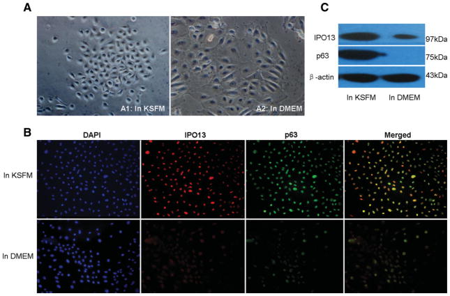
Figure 4.
TKE2 cells cultured in different media. (A): TKE2 cells cultured in keratinocyte serum-free defined medium (KSFM) had small-cell size with big nucleus/cytoplasm ratio (A1) and TKE2 cells rendered into large squamous cells when medium was switched from KSFM into Dulbecco’s modified Eagle’s medium (DMEM) supplemented with 10% fetal bovine serum (FBS) for 2 days (A2). (B): Immunofluorescent double staining for IPO13 and P63 by TKE2 cells cultured in different media. TKE2 cells in KSFM show intensive staining for IPO13 and P63 in nuclei and both of them have similar expression patterns. However, TKE2 in DMEM supplemented with FBS shows significant decreasing staining for IPO13 and P63. (C): Western blotting assay. TKE2 cells in KSFM show intensive expression for IPO13 and P63, but TKE2 cells in DMEM supplemented with FBS show downregulated expression of IPO13 and no expression of P63. Original magnification: ×200

