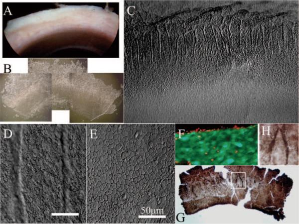
Figure 1.
Enrichment of p63-positive cells in limbal palisades. From one quarter of corneo-limbo-scleral tissue left after corneal transplantation (A), an intact limbal epithelial sheet was isolated by overnight dispase digestion (B). When one fourth of an isolated sheet (i.e., one sixteenth of the entire limbal sheet) was put on a plastic dish (C), pigmented limbal palisades noted before digestion (A) were preserved and exhibited undulating folds in the limbal location (B, C), where cells were small (D). In contrast, cells in the peripheral corneal region were flat and larger (C, E). Cells of such isolated limbal sheets were viable, as revealed by green fluorescence in a cell-viability assay, whereas some cells in the cut edge were dead, as shown by red fluorescence (F). Immunostaining showed that p63-positive cells were enriched in limbal palisades (G, enlarged inset in H). Bars, 50 μm.

