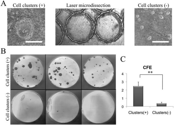
Figure 3.
Higher clonal growth by p63-positive cell clusters. When dispase-isolated human limbal sheets were cultured on plastic in SHEM for 48 hours (Fig. 2), the cells were collected from areas with (+) and without (−) p63+ clusters by a laser-assisted microdissection microscope (A) and rendered into single cells by trypsin/EDTA. After being seeded on 3T3 fibroblast feeder layers for 14 days, cells derived from p63+ cluster areas exhibited more vivid clonal growth revealed by rhodamine B staining (B) and a significantly higher CFE than those from the noncluster areas (C, **P < 0.01). Bars, 50 μm.

