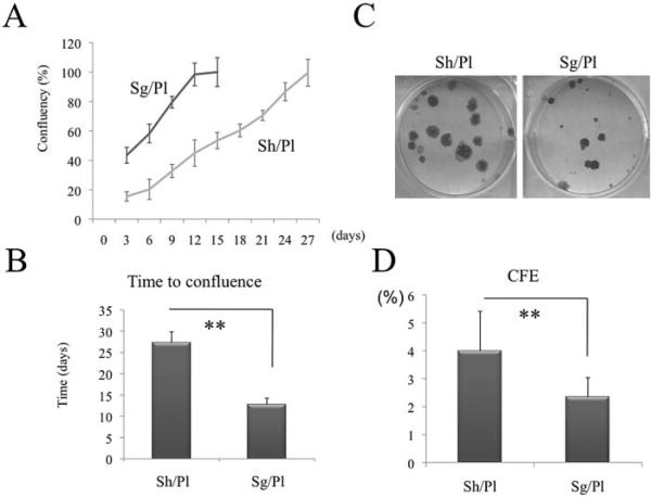
Figure 4.
Comparison of clonal growth potential between sheets and single cells. Dispase-isolated limbal epithelial sheets were cultivated Sh/Pl or Sg/Pl after trypsinization for 27 days. As expected, epithelial growth measured by digitizing phase-contrast micrographs and plotted as the percentage of confluence in Sg/Pl was significantly greater (A) and faster (B, P < 0.01) than that in Sh/Pl. Afterward, the same number of single cells obtained from the above two cultures was seeded on 3T3 fibroblast feeder layers. The resultant clonal growth by Sh/Pl was more vivid (C) and had a significantly higher CFE (D) than that by Sg/Pl (**P < 0.01).

