Figure 5.
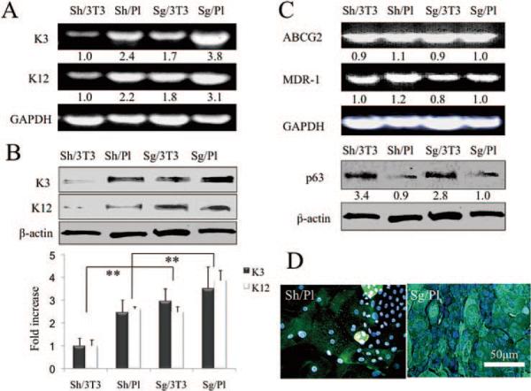
Expression of differentiation and progenitor markers. RT-PCR showed that expression of K3 and −12 transcripts (i.e., corneal differentiation markers), was the lowest in sheets expanded on 3T3 fibroblast feeder layers (Sh/3T3) when compared to the rest (A). Western blot analysis also showed that expression of K3 and −12 proteins was the lowest in Sh/3T3 (B, **P < 0.01). Although expression of ABCG2 and MDR-1 transcripts did not show any difference among the four conditions, that of p63 protein by either sheets or single cells was promoted by 3T3 feeder cells (C). Immunostaining for K3 was heterogeneously positive in sheet cultures on plastic, but was uniformly positive in single cell suspension cultures (D).

