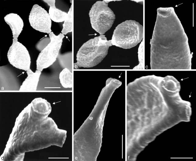
Fig. 3.
Scanning electron micrographs of Cladosporium sphaerospermum NRRL 8131. a, b. Branching chains of conidia, showing conidiogenous loci with disjunctors (arrows); c. apex of conidiophore with conidiogenous scar in profile (arrow); d. two conidiogenous loci at apex of a secondary ramoconidium, the upper (arrow) clearly coronate; e. two conidiogenous loci at apex of a conidiophore, the one facing the viewer is clearly coronate (arrow); f. two conidiogenous loci (arrows) at apex of a secondary ramoconidium are coronate. — Scale bars: a–c = 2.5 μm, d = 1 μm, e = 5 μm, f = 1.25 μm.

