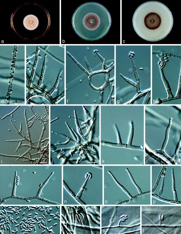
Fig. 8.
Phaeoacremonium rubrigenum. a–c. Sixteen days old colonies on 2 % MEA (a), PDA (b) and OA (c). — d–q. Aerial structures on 2 % MEA; d. mycelium showing prominent exudate droplets observed as warts; e–h. single conidiophores; i, j. branched conidiophores; k, l. type I phialide; m, n. type II phialide; o, p. type III phialide; q. conidia. — r–t. Structures on the surface of and in 2 % MEA: adelophialides with conidia; all from H-20121 (holotype); d–t: DIC. — Scale bars: d = 10 μm; scale bar for d applies to i–k and k–t.

