
Fig. 2.
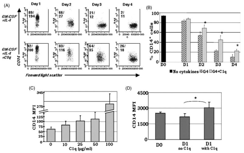
C1q sustains CD14 expression on monocyte-derived DCs in culture. MNCs isolated from PB by density gradient centrifugation were cultured in the presence of GM-CSF + IL-4 ± 25 μg/ml C1q (A,B), or without the addition of cytokines ± 25 μg/ml C1q (D). For the dose-response analysis, several concentrations of C1q were added as indicated (C). Cells were analyzed on days 0–4 for the expression of CD14. (A) Dendritic cells cultured in the presence of C1q retained CD14 on their surface and maintained elevated CD14 expression until day 4. The numbers in the upper corners of the plots represent percentage of positive cells/MFI. One representative experiment illustrated by dot plot analysis is shown. (B) Temporal analysis performed by flow cytometry revealed significantly higher levels of CD14 expression in C1q treated cultures (G4 + C1q) compared to G4 alone until day 4. *P < 0.05, **P < 0.01 (n = 4) (C) Dose-response analysis confirmed that the MFI of CD14+ cells correlates positively with increasing doses of C1q on day 2 (n = 3). (D) Baseline CD14 expression (day 0) on monocytes increased after 24 h of C1q treatment without the addition of DC growth factors (n = 3). Cells were gated on the HLA-DR+ population for all experiments.