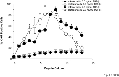
Figure 1.
Percentage of Ki-67–positive cells at 0 and 2 ng/mL TGF-β1. At 0 ng/mL TGF-β1, there are small and similar percentages of anterior and posterior cells entering the cell cycle. At 2 ng/mL TGF-β1, there is a greater percentage of posterior cells staining positive for Ki-67 at earlier time points (<7 days). At this same concentration, at later time points (greater than 7 days), there is a greater percentage of anterior cells staining positive for Ki-67.

