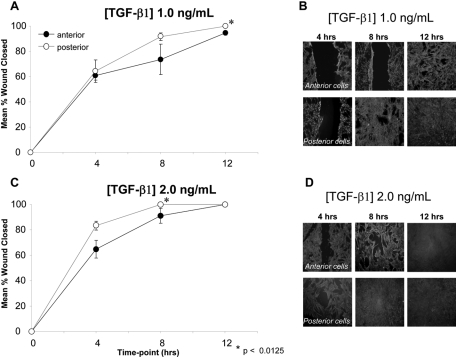
Figure 5.
Closure of a mechanically induced wound in cultured anterior and posterior cells. (A) At TGF-β1 concentration of 1 ng/mL, posterior cells demonstrate faster wound closure than anterior cells over a 12-hour period. (B) Photomicrographs of α-SMA–stained wounded cell cultures exposed to 1 ng/mL TGF-β1. Note the consistently larger wound area exhibited by anterior cells relative to their posterior counterparts at any given postscratch time point. (C) At a TGF-β1 concentration of 2 ng/mL, posterior cells again demonstrate faster wound closure than anterior cells. (D) Photomicrographs of α-SMA–stained wounded cell cultures exposed to 2 ng/mL TGF-β1. Note the consistently larger wound area exhibited by anterior cells relative to their posterior counterparts at each time point after wounding.

